Super Resolution Microscope N-SIM
3D-SIM mode
Comparison of images captured with an N-SIM and a conventional confocal microscope
E. coli (XL1-Blue) expressing SGFP2
Photos courtesy of: Drs. Takahisa Suzuki and Ikuo Wada, Fukushima Medical University School of Medicine
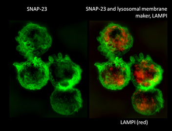
Macrophages (J774 cells expressing mVenus-SNAP23) labeled with Alexa488 and Alexa594, and immunostained with GFP and LAMPI antibodies after fixation.
Photos courtesy of: Drs. Chiye Sakurai, Kiyotaka Hatsuzawa and Ikuo Wada, Fukushima Medical University School of Medicine
LLC-PK1 cells
eEmerald to Lif Act
Photo courtesy of: Mike Davidson, Florida State University

Luminal surface of the organ of Corti at postnatal day 1. (Mouse)
Green: F-actin.
Red: Acetylated Tubulin
Specimen courtesy of: Ms. Kanoko Kominami, Dr. Hideru Togashi and Prof. Yoshimi Takai, Division of Molecular and Cellular Biology, Kobe University Graduate School of Medicine
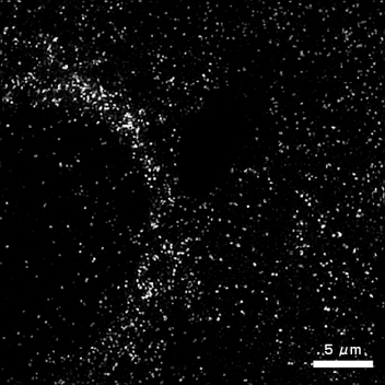
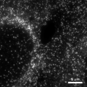
TIRF-SIM mode
Comparison of images acquired with TIRF-SIM and with conventional TIRF
With TIRF-SIM
4-hour time-lapse with images every minute
With conventional TIRF
FoLu (fox lung) cells
eGFP Vinculin
Photos courtesy of: Mike Davidson, Florida State University
With TIRF-SIM
With conventional TIRF
BSC-1 cells, African green monkey kidney epithelial
Alexa Fluor-568 Clathrin
Photos courtesy of: Mike Davidson, Florida State University