Super Resolution Microscope N-SIM
Temporal resolution of 0.6 sec./frame enables super resolution time-lapse imaging of dynamic live cell events

In Structured Illumination Microscopy, the unknown cellular ultra-structure is elucidated by analyzing the moiré pattern produced when illuminating the specimen with a known high-frequency patterned illumination. Nikon's Structured Illumination Microscopy (N-SIM) realizes super resolution of up to 85nm in multiple colors. In addition, it can continuously capture super resolution images at temporal resolution of 0.6 sec./frame, enabling the study of dynamic interactions in living cells.
Live cell imaging at double (to approx. 85nm) the resolution of conventional optical microscope
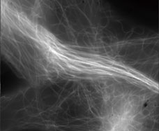
The N-SIM super resolution microscope utilizes Nikon's innovative new approach to "Structured Illumination Microscopy" technology. By pairing this powerful technology with Nikon's renowned CFI Apo TIRF 100x oil objective lens (NA 1.49), N-SIM nearly doubles (to approx. 85nm*) the spatial resolution of conventional optical microscopes, and enables detailed visualization of the minute intracellular structures and their interactive functions.
- *Excited with 488nm laser, in TIRF-SIM mode
- Microtubules in B16 melanoma cell labeled with YFP
Objective: CFI Apo TIRF 100x oil (NA 1.49) Image capturing speed: approx. 1.8 sec./frame (movie)
Photographed with the cooperation of: Yasushi Okada, Ph.D., Department of Cell Biology and Anatomy, Graduate School of Medicine, University of Tokyo
With conventional microscope

With N-SIM (3D-SIM mode)
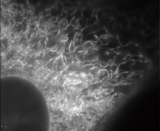
- Endoplasmic reticulum (ER) in living HeLa cell labeled with GFP
Objective: CFI Apo TIRF 100x oil (NA 1.49) Image capturing speed: approx. 1.5 sec./frame (movie)
Photographed with the cooperation of: Ikuo Wada, Ph.D., Institute of Biomedical Sciences, Fukushima Medical University School of Medicine
With conventional microscope

With N-SIM (3D-SIM mode)
Temporal resolution of 0.6 sec./frame–amazingly fast super resolution microscope system
N-SIM provides ultra fast imaging capability for Structured Illumination techniques, with a time resolution of up to 0.6 sec/frame, which is effective for live-cell imaging (with TIRF-SIM/2D-SIM mode; imaging of up to approx. 1 sec./frame is possible with 3D-SIM mode).
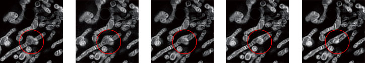
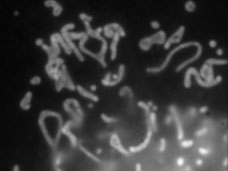
- Dynamics of mitochondria stained with Mito-Tracker red
Cristae in mitochondria are visualized and the dynamics of mitochondria can be observed.
Mode: 3D-SIM
Objective: CFI Apo TIRF 100x oil (NA 1.49)
Image capturing interval: approx. 1 sec. (movie)
Various observation modes
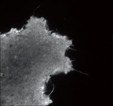
TIRF-SIM/2D-SIM mode
This mode captures super resolution 2D images at high speed with incredible contrast. TIRF-SIM takes advantage of Total Internal Reflection Fluorescence observation at double the resolution as compared to conventional TIRF microscopes, facilitating a greater understanding of molecular interactions at the cell surface.
- Plasma membrane of B16 melanoma cell labeled with YFP
Objective: CFI Apo TIRF 100x oil (NA 1.49)
Photographed with the cooperation of: Yasushi Okada, Ph.D., Department of Cell Biology and Anatomy, Graduate School of Medicine, University of Tokyo
Conventional
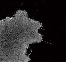
TIRF-SIM
3D SIM capable
Axial super resolution observation using the N-SIM system enables optical sectioning of specimens at 300nm resolution in cells and tissues of up to 20µm thickness. Additionally 3D SIM eliminates out of focus background fluorescence resulting in breathtaking contrast.
- Mitochondria of NIH-3T3 cell labeling MitoTrackerRed. The Crista is observed clearly by N-SIM
Conventional

3D-SIM
5 laser multi-color super resolution capability
The Nikon LU-5 is a modular system with up to 5 lasers enabling true multi-spectral super resolution. Multi-spectral capability is essential to the study of dynamic interactions of multiple proteins of interest at the molecular level.

Co-localization images of a target protein of VGEF signaling (Cy3) and its ubiquitin E3 ligase (FITC)
Unprecedentedly detailed structure of the nuclear body can be observed
Mode: 3D-SIM, Z-stack
Objective: CFI Apo TIRF 100x oil (NA 1.49)
Photographed with the cooperation of: Hidetaka Ohnuki, Ph.D., Shigeki Higashiyama, Ph.D., Ehime University Graduate School of Medicine
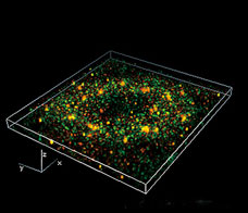
3D reconstruction image approx. 5µm thick (part)
The Principle of the Structured Illumination Microscopy
Analytical processing of recorded moiré patterns produced by overlay of a known high spatial frequency pattern mathematically restores sub-resolution structure of a specimen.
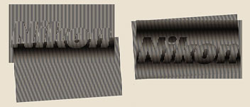
Irradiation of striped pattern illumination with known spatial frequency allows acquisition of information from a minute structure as moiré fringes.
Utilization of high spatial frequency laser interference to illuminate sub-resolution structure within a specimen produces moiré fringes, which are captured. These moiré fringes include modulated information of the sub-resolution structure of the specimen.
Through image processing, the unknown specimen information can be recovered to achieve resolution beyond the limit of conventional optical microscopes.
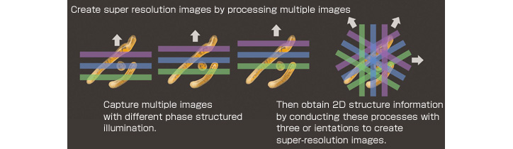
Create super resolution images by processing multiple moiré pattern images
An image of moiré patterns captured in this process includes information of the minute structures within a specimen. Multiple phases and orientations of structured illumination are captured, and the displaced "super resolution" information is extracted from moiré fringe information. This information is combined mathematically in "Fourier" or aperture space then transformed back into image space creating an image at double the conventional resolution limit.
Utilizing High Frequency striped illumination to double the resolution
The capture of high resolution, high spatial frequency information is limited by the Numerical Aperture (NA) of the objectives, and spatial frequencies of structure beyond the optical system aperture are excluded (Fig. A). Illuminating the specimen with high frequency structured illumination, which is multiplied by the unknown structure in the specimen beyond the classical resolution limit, brings the displaced "super resolution" information within the optical system aperture (Fig. B).
When this "super resolution" information is then mathematically combined with the standard information captured by the objective lens, it results in an effective doubling of the NA, and therefore resolution of the optical system (Fig. C).
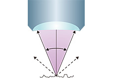
Fig. A: Resolution is limited by the NA of the objective

Fig. B: The product of Structured Illumination and normally unresolvable
specimen structure produce recordable moiré fringes containing the specimen information at double the conventional resolution limit.
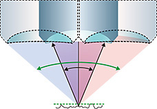
Fig. C: Rays with approx. double the angle of the NA are captured