Confocal Optical System
Fig. 1: Confocal optical system
A confocal optical system is an optical system that determines reflectance of light from a small targeted spot at a single focal point. The system’s pinhole aperture restricts the degree of reflected light from the sample in order to keep unwanted optical light from interfering with focus on the surface of the sample. Because of this reason, a confocal optical system helps make images clear and well contrasted.
Fig. 1 illustrates how the confocal optical system works. Only light that has been transmitted from the light source and has passed through the pinhole aperture can emerge, go forward through the objective lens, reflect from the sample and return. In this case, only that light focused at the focal point of the sample can form an image at the pinhole aperture and advance (see red line). The light, which cannot create an image at the surface of the sample (indicated by blue line), cannot pass through the pinhole aperture. It is therefore possible to obtain a clear image by using a detector to catch only the reflected light that has passed through the pinhole aperture.

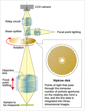
Fig. 2: Nikon’s original optical system
Now, let’s take a look at the confocal optical system in Nikon’s CNC video measuring system. Designed to automatically measure dimensions and shapes of various precision parts and electronic components, its workings are illustrated in Fig. 2.
Fig. 3 shows an example of optical microscope observation of a fine detail on an IC board. Try to measure the semispherical shape (Fig. 3) using an optical microscope, and black-and-white pictures appear on the display, one by one, as measurement progresses. As the objective lens is moved slowly toward the object to be measured, the region of the object closest to the lens is in focus at first, and the light reflected from that region returns through the pinhole aperture. This light is captured by a CCD camera. As the objective lens is lowered, the light toward the outline of the semisphere returns through the aperture. As the objective lens is further lowered, the light at the outer edge of the semisphere returns through the aperture. In other words, the sample is not shot at once by a camera; separate regions of the sample toward which light is emitted are various “sliced” perspectives of the targeted sample, and these sliced perspectives are processed by computer into a three-dimensional image.
A single, fast, one-second vertical scan determines each pixel’s height. Respective case in which pixel A or pixel B is brightest (in focus) can be obtained when summarizing data. By comparing data indicating differences between focal point positions, sample height can be calculated on a nanometer scale. The color photo shown was created by color-processing combined data. Red and blue coloration distinguishes high and low regions, respectively.
Fig. 3: Three-dimensional image construction process
A disk called a Nipkow disk provides an immense number of pinhole apertures and rotates at high speed. The confocal optical system emits pinpoint light and catches that light returning from an object. It is possible to scan the entire object by emitting light through a rotating Nipkow disk. The Nipkow disk is made by applying detection expertise that takes number and arrangement of pinhole apertures precisely into account.
The confocal optical system, which is particularly excellent for height observation, is widely used in biomicroscopes as well as measuring instruments. It is indispensable for observing the inside of samples like cells without compromising focus.
Posted August 2007
