Cell Culture Observation SystemBioStation CT
Stem cell screening inside the incubator
With conventional cell monitoring procedures, a culture vessel has to be taken out of the incubator for microscope observation, where cells are subjected to stressful environmental changes and vibration. Researchers then have to spend additional time repositioning the vessel to find the same observation points. Nikon's BioStation CT eliminates these problems by providing a stable environment so that the cultures don't suffer while they are being imaged and allowing for a complete trace of the same live cells, including stem cells.
Advanced basic functions
Automatic image capture

The autofocus mechanism allows the capture of in-focus images. Z-stack imaging in phase contrast observation, multi-sample imaging and multi-point imaging are possible with multiple magnifications. User-configured imaging conditions that can be saved in BioStation CT support the repeatability of observations.
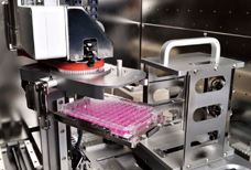
Automatic vessel transportation
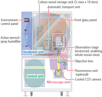
BioStation CT incorporates a transport unit that provides stable vessel transportation within the heated and humidified incubation area. The high-precision motorized stage in the observation unit allows for automated imaging of the entire area of a well in all culturing formats.
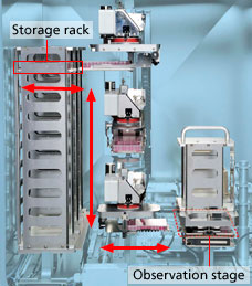
The transport unit carefully conveys the vessels from the storage rack to the observation stage in accordance with configured schedules.
Remote access

Configuring the imaging settings, scheduling a time-lapse experiment, and viewing the cell images are possible via a network. The captured data can be automatically downloaded to the user's local computer. This enables users to monitor the cell status away from the laboratory. When a culture environment (temperature, humidity, CO2 concentration) control error occurs, BioStation CT can notify the users of the error by e-mails.
Various functions
Full-well scan imaging and highly magnified image stitching
High-resolution full-well scans are reconstructed by stitching captured adjacent images. This enables clear detection of an iPS colony, which is difficult to detect because of its low induction efficiency, no matter where it forms in the vessel. The specified position of the vessel can be highly magnified with high resolution. BioStation CT also offers cell registration to allow for repeated visits to the same location. These time-lapse sequences can be created even when a vessel is removed from the BioStation CT for medium exchange.
- [Note] Depending on the vessel used, the BioStation CT may not be able to focus on some areas.
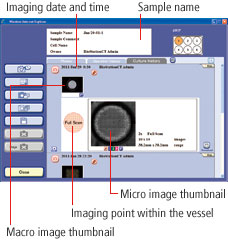
Micro observation
Phase contrast and fluorescence images can be captured with the high-sensitivity cooled CCD camera. These images can be magnified in 2x, 4x, 10x, 20x and 40x. Up to 40 phase contrast images can be captured along the Z axis with the Z-stack function.

Macro observation
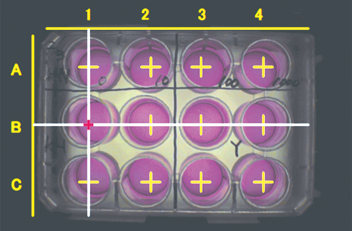
Brightfield image of the whole vessel provides users outside the BioStation CT with information such as handwritten information on the vessel, medium color and whether mold is growing or not. In addition, alkaline phosphatase stained cell counting is possible with the optional image analysis software CL-Quant.
Stable culture environment maintenance
Precise temperature control
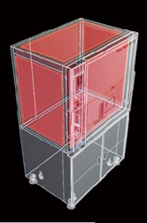
The inside temperature is directly controlled by panel heaters embedded in the incubator's six sides. This allows highly precise temperature maintenance.
Humidity control with air-flow type active aerosol spray humidifier

Distilled water is automatically sprayed inside the incubator to keep the optimum humidity. Water can be supplied to the tank without opening the incubator door. This air-flow type humidifier reduces contamination risks compared to the water bath type.
Environment data recording
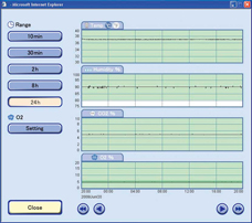
The culture environment is constantly monitored and recorded. The environment data can be accessed at anytime.
Smooth vessel transportation
The waver of liquid surface during the transportation is less than 2 mm. The drift and stress of cells are reduced.
Reduced contamination risk
The incubator interior can be sterilized using hydrogen peroxide gas. (This is optional, and a 200 V power source is necessary.)
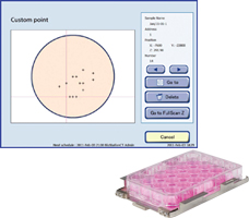
Different GUI for each vessel type
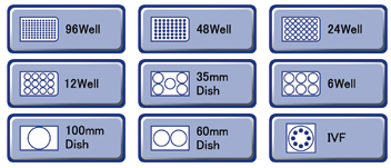
Vessel type icons
Wells to be observed can be chosen on the touchscreen.
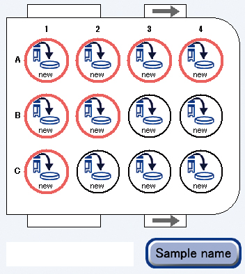
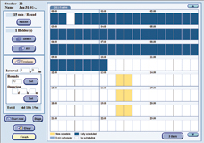
GUI for 12-well plate
Easy operations
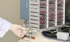
1. Vessel installation
Culture vessel installation into the storage rack
Efficient installation with the optional sliding storage rack that enables multiple vessels to be installed at one time
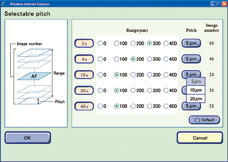
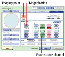
2. Imaging parameter setting
Easy touchscreen operation
Time-lapse imaging configurations such as magnification, imaging point, fluorescence channel and stage motion speed can be set.
3. Scheduling
Time-lapse imaging schedule
The imaging interval and total period can be set. The shortest time-lapse imaging interval is one minute.
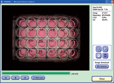
4. Captured image view
Culture history data management
The time-course change of a specimen can be observed easily in sequentially displayed captured images.
5. Medium exchange
High-precision repeatability
Accurate tracing of same cells, even after medium exchange, is possible using a dedicated tray holder, as BioStation CT records culture history, such as medium exchange and subculture, as well as X-Y positions for each vessel.
6. Data report
Reliable data management and documentation support
Obtained data is duplicated and protected using uninterruptible power supply. Observation information such as temperature, humidity and imaging date can be written and displayed on the captured image to simplify presentation document preparation.

-
-
Nikon will exhibit at the ISSCR 11th Annual Meeting. BioStation CT will be on display.
-
 Stem Cell Research – Nikon BioStation CT