
![]()
The ISSCR's annual meeting has become the world's premier stem cell research event. The meeting serves as the largest forum for stem cell and regenerative medicine professionals from around the world. Nikon will present products and applications at the event. It will also offer the presentation Innovation Showcase.

-

-
- SPEAKER:
Professor Lee L. Rubin, PhD -
Director of HSCI's Therapeutic Screening Center Professor in Harvard University, USA
- SPEAKER:
- DATE: Friday, June 14
- TIME: 11:45 a.m. – 12:15 p.m.
- VENUE: Room 258 A-C
-
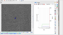
Nikon BioStation CT realizes long term time-lapse imaging and advanced analysis not only on fluorescent images but also Phase contrast images.
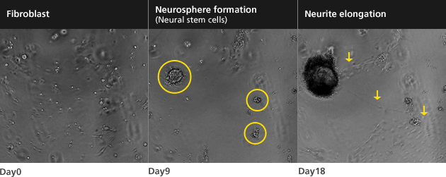
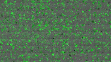
- Imaging of the direct induction from mouse fibroblasts to Neural stem cells and neurons
-
Stem Cells. 2012 Jun;30(6) : 1109-19
- Courtesy
Professor Hideyuki Okano and Dr. Takeshi Matsui Department of Physiology, Keio University School of Medicine - Magnification : 4X
- Culture period : 18 days
- Imaging interval : 4 hours
- Courtesy
-
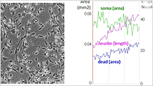
- Neural differentiation analysis
-
- Typical iPS colony formation movie
-
- Imaging of reprogramming process
-
- Human iPS colonies prediction within first 72 hours of colony formation
Nikon BioStation CT is an advanced, automated cell culture observation instrument specialized for stem cell research.
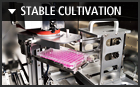
Ideally suited for live cell imaging, BioStation CT maintains a stable environment during microscopic image acquisition.
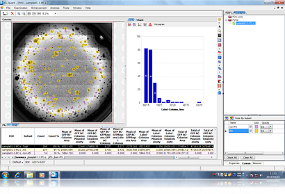
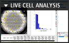
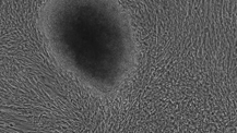
Whole well scan imaging enables clear detection of an iPS colony, which is difficult to detect because of its low induction efficiency. Combination with advanced image analysis technology enable to be revolutionary new approach and lead to improvements in cell characterization methodologies and a better understanding of the reprogramming and differentiation process. Imaging and analysis become processed automatically by separate PC with no stress. The left Figure shows the result of automated classification into iPS and non-iPS cell colonies from the phase contrast images. (The entire area of a 100mm dish)
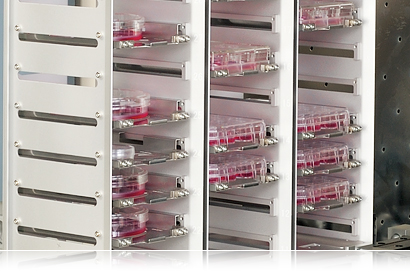
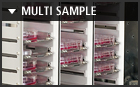
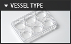
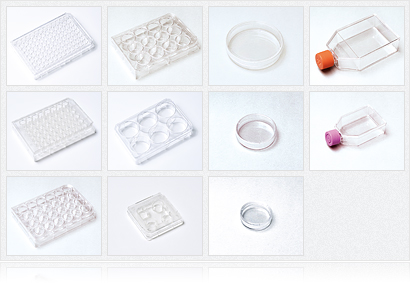
BioStation CT can incubate total 30 vessels in case of well plate at once. Researchers can examine the differences in each culture vessels.
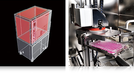
Temperature, Humidity, CO2 concentration and O2 concentration are controlled by accurate sensor and feedback system.
- Harvard Stem cell Institute
- Morgridge Institute
- Diabetes Center
- UCR Stem Cell Core Facility
- USC Stem Cell Core
- Newcastle University
- University Hospital Bern
- Gurdon Institute
- Masaryk University
- China Agricultural University
- Kyoto University
- Keio University
- National Institute of Biomedical
- Innovation
- Neural Stem Cells Directly Differentiated from Partially Reprogrammed Fibroblasts Rapidly Acquire Gliogenic Competency
-
Takeshi Matsui, Morito Takano, Kenji Yoshida, Soichiro Ono, Chikako Fujisaki, Yumi Matsuzaki, Yoshiaki Toyama, Masaya Nakamura, Hideyuki Okano, Wado Akamatsu
Stem Cells. 2012 Jun;30(6):1109-19 - Protein Kinase C Regulates Human Pluripotent Stem Cell Self-Renewal
-
Masaki Kinehara, Suguru Kawamura, Daiki Tateyama, Mika Suga, Hiroko Matsumura, Sumiyo Mimura, Noriko Hirayama, Mitsuhi Hirata, Kozue Uchio-Yamada, Arihiro Kohara, Kana Yanagihara, Miho K. Furue
PLOS ONE. January 2013 | Volume 8 | Issue 1 | e54122
- Generation of hyaline cartilaginous tissue from mouse adult dermal fibroblast culture by defined factors
-
Kunihiko Hiramatsu, Satoru Sasagawa, Hidetatsu Outani, Kanako Nakagawa, Hideki Yoshikawa, and Noriyuki Tsumaki
J Clin Invest. 2011;121(2):640–657 - Efficient and Reproducible Myogenic Differentiation from Human iPS Cells: Prospects for Modeling Miyoshi Myopathy In Vitro
-
Akihito Tanaka, Knut Woltjen, Katsuya Miyake, Akitsu Hotta, Makoto Ikeya, Takuya Yamamoto, Tokiko Nishino, Emi Shoji, Atsuko Sehara-Fujisawa, Yasuko Manabe, Nobuharu Fujii, Kazunori Hanaoka, Takumi Era, Satoshi Yamashita, Ken-ichi Isobe, En Kimura, Hidetoshi Sakurai
PLOS ONE. April 2013 | Volume 8 | Issue 4 | e61540

-
- Nikon Instruments Inc.
-
1300 Walt Whitman Road, Melville, N.Y. 11747-3064, U.S.A.
Phone: +1-631-547-8500 : +1-800-52-NIKON (within the U.S.A. only)
Fax: +1-631-547-0306
E-mail: nikoninstruments@nikon.net

-
- Nikon Instruments Europe B.V.
-
Tripolis 100, Burgerweeshuispad 101, 1076 ER Amsterdam, The Netherlands
Phone: +31-20-7099-000
Fax: +31-20-7099-298

-
- Nikon Corporation
-
Shin-Yurakucho Bldg., 12-1, Yurakucho 1-chome, Chiyoda-ku, Tokyo 100-8331 Japan
Phone: +81-3-3216-2375
Fax: +81-3-3216-2385
E-mail: Support.Biostation@nikon.com - Nikon Instruments (Shanghai) Co., Ltd.
-
26F, Hang Seng Bank Tower, No.1000 Lujiazui Ring Road, Pudong New District, Shanghai 200120, China
Phone: +86-21-6841-2050
Fax: +86-21-6841-2060
Can't find your country or area? Please visit Worldwide Network for your local contact information.