Time Lapse Imaging System BioStation IM-Q
- The perfect solution for long-term, time-lapse, live-cell imaging
- Simple and easy operation
- Two kinds of analysis software are available for intended use. (Option)
- Optional accessories broaden the range of applications

The perfect solution for long-term, time-lapse, live-cell imaging
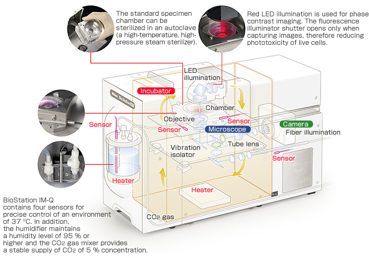
The BioStation IM-Q incorporates a microscope, an incubator and a high-sensitivity cooled CCD camera in a compact body. This all-in-one package provides a stable environment for live cells and advanced solutions for simple long-term time-lapse data acquisition.
The BioStation IM-Q eliminates the need for a darkroom for fluorescence imaging, meaning it can be installed anywhere.
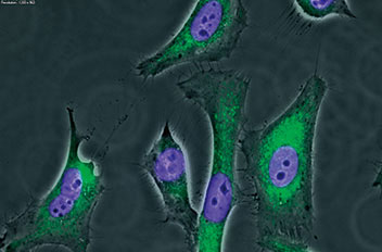
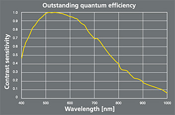
Exceptional high-sensitivity fluorescence images
The high sensitivity of the built-in CCD camera, equivalent to ISO800, reduces exposure time, which minimizes photobleaching and damage to specimens while increasing throughput for multipoint acquisition. The cooling mechanism prevents heat-induced noise and allows even weak fluorescence to be captured. The fluorescence equipment employs a noise terminator mechanism in order to eliminate stray light. This enables high-contrast imaging with high S/N ratios.
Cell-friendly environmental control
Cell culture and image capture functions are beautifully integrated. No complex setup or alignment procedures that conventional time-lapse observation systems require are necessary. The vibration-proof and heat-insulated structure enables stable image acquisition even over long periods.
Accurate recording of live-cell dynamics over a number of days
Focus drift during lengthy time-lapse observations has been greatly reduced, enabling reliable time-lapse imaging even for several days. The temperature of the chamber, the chamber exterior and microscope unit is precisely controlled with built-in heaters and fans. The focusing mechanism is made from thermal-stable materials and is therefore resistant to thermal expansion. Moreover, BioStation IM-Q moves the objective lens instead of the stage. This eliminates focus drift caused by vibration of culture dish, minimizing stress on the cells.
Lengthy time-lapse imaging without focus drift
Membrane-anchored GFP of rat hippocampal neurons was captured in 120-hour time-lapse imaging.
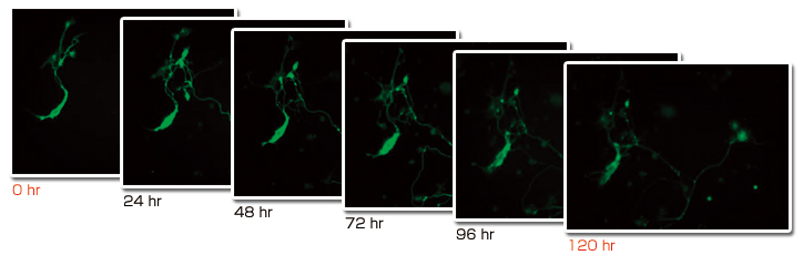
Photos courtesy of: Dr. Chieko Nakada, Dr. Yuuri Nemoto, Dr. Hiroko Hijikata, Dr. Akihiro Kusumi, Institute for Integrated Cell-Material Sciences. Kyoto University
Simple and easy operation
Operation from a PC
BioStation IM-Q provides fully motorized control from a PC, allowing users who are not accustomed to operating a microscope or camera to easily conduct time-lapse imaging. The user only has to set the culture dish in place and program the image recording.
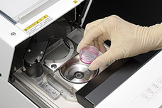
1. Culture dish setup
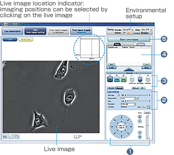
2. Setting with intuitive GUI
- Set live image location with XYZ movement control.
- Configure imaging condition.
- Select illuminations, filters and magnifications.
- Input imaging time interval.
- Start time-lapse imaging.

3. Time-lapse imaging and analysis
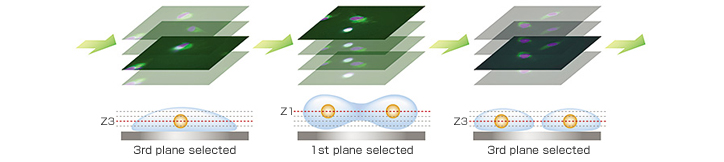
Image file output at desired Z-axis planes
Images at different Z-axis planes can be selected from the captured Z-stack images at each time point and assembled into a seamless movie file. This is optimal for imaging a specimen in which the observation point moves along the Z-axis direction, such as with cell division.
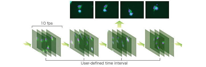
Streaming function
Rapid motion changes such as cardiomyocyte beats are captured by high-speed 10-fps imaging at user-defined time intervals.
Ergo controller
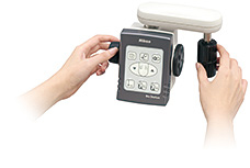
An ergo controller allows X, Y and Z directional movement with an operational feel similar to a microscope. It also allows changeover of magnifications, fluorescence filters, imaging/observation methods and fluorescence shutter ON/OFF.
Widefield display
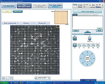
Because a wide 6 mm x 6 mm area can be displayed by image stitching, the point of time-lapse observation can be easily specified from the widefield image.
Two kinds of analysis software are available for intended use. (Option)
Imaging software NIS-Elements Ar
Nikon's proprietary imaging software NIS-Elements Ar allows multi-dimensional image capture, image processing, and data management and analysis of up to 6D.
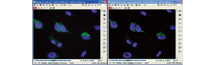
Deconvolution
Haze and blur of an image that can occur when capturing a thick specimen or a fluorescence image can be eliminated from the captured image.
Cell counting
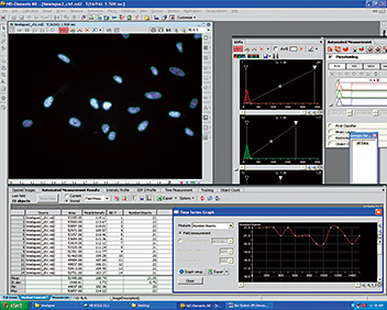
Images are processed by image analysis routines and the extracted objects can be counted.
Slice view

Images in three orthogonal planes (sliced images along the XY-, YZ-, and ZX-planes) can be viewed in a single display.
Volume rendering
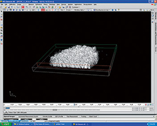
3D images can be reconstructed from captured Z-stack images.
Dedicated image analysis software for BioStation series-CL-Quant
Cell detection in phase contrast images
CL-Quant automatically detects and measures the cellular area in unstained, label-free phase contrast images. Unique image processing algorithms provide accurate thresholding of phase contrast images, which enables non-invasive quantitative analysis of cells. Cell detection accuracy can be improved through a learning function process.
Analysis option: cell counting, scratch (wound) test, growth curve, tracking, stem cell colony analysis
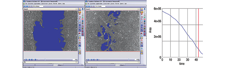
- Scratch (wound) test quantification
- CL-Quant automatically segmented the acellular area (blue) in the time-lapse cell migration images that were captured at the beginning (left) and on the second day (right) of a 2-day phase contrast observation. The graph indicates how the acellular area decreased.
Optional accessories broaden the range of applications
Motorized chamber for four-well culture dish
Automated changeover allows each of the four culture wells to be imaged. Observation of four different samples is possible in a single time-lapse experiment, facilitating comparative analyses.
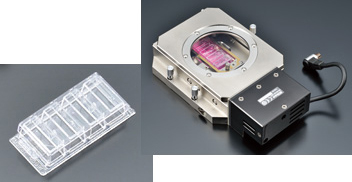
Lab—Tek™chambered coverglass
(Cat. No. 155383. Nunc™)

Operation screen
Specialized Hi-Q4 culture dish that enables multi-sample observation
This new 35 mm culture dish is divided into four parts and has an incorporated plane parallel top plate. The plate prevents light path distortion by the meniscus, which is the curve at the air-water interface, and enables high-quality phase contrast observation.
- *Use of the Hi-Q4 culture dish is available for the BioStation IM-Q CELL-S2 unit only.
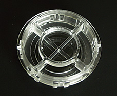
Hi-Q4 dish
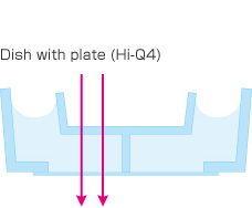
The absence of a meniscus allows light to pass through without refrection
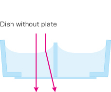
Light refracted by the media's meniscus distortion and poor phase image.
Perfusion components
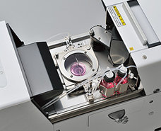
The perfusion components allow reagent administration and medium exchange without a change to the culture environment.
Two—bottle installation
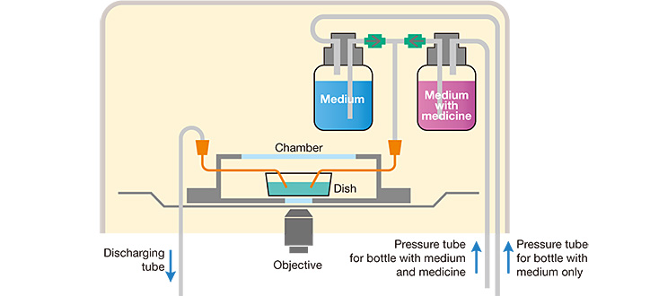
- *Syringe pump or peristaltic pump is necessary to pressurize the solution bottles.