
GaAsP multi detector unit
Nikon developed the GaAsP multi-detector unit equipped with two GaAsP PMTs and two normal PMTs.
A GaAsP PMT has much higher sensitivity than a normal PMT, thus acquisition of brighter signals with minimal background noise is possible with a GaAsP PMT, even with weak fluorescence, which, until now, has been difficult to detect.
When using resonant scanners, the GaAsP PMT enables low-noise, high-speed imaging. (See graphic below titled “2. Image comparison” to compare images acquired using a resonant scanner).
- Comparison images are acquired with the same excitation laser power and scanning speeds, in order to compare the difference in PMT sensitivity. Adjusted PMT gain is used to achieve the same fluorescence intensity.
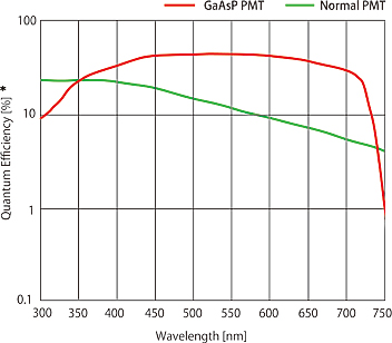 Sensitivity comparison of GaAsP PMT and normal PMT
GaAsP PMT realizes higher sensitivity than normal PMT, thus offering high quantum efficiency up to 45%.
- * Quantum efficiency indicates logarithm
1. Image comparison of GaAsP PMT and Normal PMT (acquired using galvano scanner)
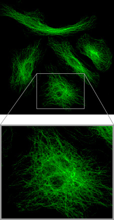 GaAsP PMT
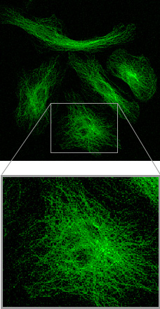 Normal PMT
Microtubule labeled with Alexa488
Background noise in the image captured using GaAsP PMT is less than that of a normal PMT, thus the minute structures of the sample are easily distinguishable.
Specimen courtesy of: Dr. Tadashi Karashima, Department of Dermatology, Kurume University School of Medicine
2. Image comparison of GaAsP PMT and Normal PMT (acquired using resonant scanner)
Images of HeLa cell labeled with MitoTracker and their intensity surface plots
Fluorescence intensity is indicated with color and height in the 3D graph.
There are major differences between GaAsP PMT and normal PMT in the background noise of the image and its temporal change.
3. Simultaneous two-color image using GaAsP PMT (acquired using resonant scanner)
Cardiomyocyte with mitochondria stained with TMRM
Calcium indicator: Fluo-8
Cardiomyocyte contraction and calcium signaling dynamics are simultaneously acquired using two-channel GaAsP PMT.
- *This movie clip is a loop sequence which repeats the recording of calcium sparks three times.