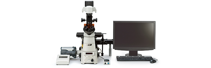
TIRF (Total Internal Reflection Fluorescence) Microscope White-light TIRF System (Multi-Fluorescence Imaging System)
Reveals vesicle fusion with high S/N ratios
One white-light illuminator supports various types of fluorescence observations (Patent pending)
The white-light TIRF system enables TIRF microscopy using mercury lamps. By exciting a confined depth, TIRF enables imaging of fluorescence images with a much higher S/N ratio than is possible using the epi-fluorescence method. The integration of a TIRF system and epi-fluorescence system enables the use of:
- White-light TIRF,
- Oblique illumination fluorescence*,
- Epi-fluorescence, and
- SRIC methods.
All modes use the same light source and switching them is simple.
- *Increasing the angle of incident light to slightly more than that of TIRF allows a deeper range of observation in the area near the coverglass.
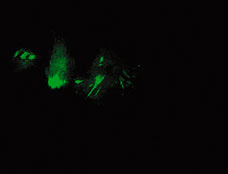
Clathrin coated vesicles images

Epi-fluorescence image

White-light TIRF image
- Specimen: GFP-tagged clathrin expressed in COS cells
- Images courtesy of Daniel Axelrod Ph,D., University of Michigan and Stephen Ross, Ph,D
Easy multiple-wavelength imaging
As this system uses a light source with a broad wavelength range, such as mercury illumination, by simply switching filters, TIRF observations are possible at a variety of wavelengths.
Evanescent Wave Illumination method
Nikon's high NA TIRF objectives make it possible to introduce laser illumination at incident angles greater than the critical angle (θc) resulting in TIRF that creates an evanescent wave immediately adjacent to the coverglass-specimen interface. The evanescent wave reaches maximally a few hundred nanometers into the specimen and its energy drops off exponentially. Nikon's laser TIRF system utilizes this evanescent wave to excite single molecules in the thin section in contact with the coverglass. Because the specimen is not excited beyond the evanescent wave, this imaging system can produce fluorescence images with an extremely high signal-to-noise (S/N) ratio.
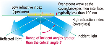
Overview of Evanescent Wave Illumination

CFI Apochromat TIRF series objectives
Numerical apertures of the highest level

These objectives are the world's first oil-immersion-type objectives to incorporate a correction ring for temperature changes and coverglass thickness. By rotating its correction collar, researchers can easily eliminate spherical aberrations' negative influence on the image quality resulting from temperature-induced changes in the refractive index of the immersion oil and influence from variation in the coverglass thickness. The lenses have been calibrated for a range from 23°C (room temperature) to 37°C (physiological temperature). Additionally, these objectives also provide spectacular images under DIC, epi-fluorescence, confocal, and deconvolution imaging, while providing a strong trapping power during applications using laser tweezers.
SRIC (Surface Reflective Interference Contrast) method can reveal focal contracts prior to switching to TIRF
As evanescent wave illumination excites within approximately 100 nm from the glass surface, TIRF observations require that specimens contact the coverglass, otherwise no TIRF image is obtained. SRIC makes all sections in contact with glass appear black, allowing users to confirm whether a specimen has adhered to the glass before proceeding with TIRF observation. As no excitation light is used in this process, specimen damage is minimized and users can take their time focusing.
Nikon has developed an SRIC system that can be used with both the laser TIRF and white-light TIRF systems. Using SRIC is as simple as switching to the special filter cube.
Laser TIRF image
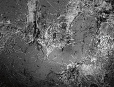
SRIC image
- Images courtesy of Dr. Masaya Hashido, University of Tokyo