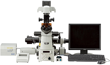
TIRF (Total Internal Reflection Fluorescence) Microscope Laser TIRF System (motorized/manual)
Enables single molecule visualization, allowing dynamic observation and functional analyses of both in vitro and living cells

Innovative motorized laser TIRF system
Newly developed motorized laser TIRF illumination unit allows laser incident angle adjustment, shutter control and switching to widefield fluorescence excitation with the control pad or NIS-Elements software. The laser incident angle can be stored with a single touch of the control pad button. Stored laser incident angles can be easily reproduced. This enables alternate time-lapse recording between fluorescence and multi-wavelength TIRF images.
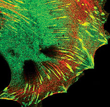
Ultra-high signal/noise ratio enables observations of single molecules
The extremely high S/N ratio created by Nikon's laser TIRF imaging system makes it possible to observe single molecules. Thanks to Nikon's proprietary Noise Terminator mechanism, the system can also produce breathtaking epi-fluorescence images with a high S/N ratio.
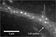
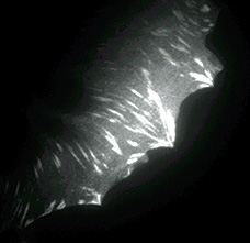
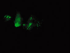
Fluorescent receptor images
Normal distribution
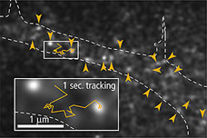
Molecular dynamics
- Specimen: YFP-tagged neurotransmitter receptors expressed in dispersed hippocampal neurons in primary culture
- Images courtesy of : Dr. Chieko Nakada, Kyoto University, Co-researcher: Professor Shigeo Okabe, Tokyo Medical and Dental University
- Bright images up to the edge of the view field are obtained.
- High-performance fluorescence cassette holder eliminates the possibility of registration shift in the optical axis for each laser wavelength.
- Simultaneous mounting of other modules such as laser tweezers is possible thanks to the expandable stratum structure of the Ti series microscope.
TIRF-C2+ System (Multimode Imaging System)
Multimode, multiangle viewing of the same field
The TIRF-C2+ system allows multimode imaging with laser TIRF, confocal and epi-fluorescence. This configuration can be a powerful tool for research, allowing the investigation of an event at the cell membrane, and the ability to follow the subsequent event cascade deep into the cells interior.
Confocal image
TIRF image

SRIC image
- Images courtesy of Shuichi Obata Ph, D., Kitasato University
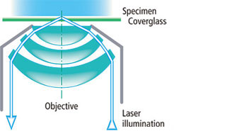
Evanescent Wave Illumination method
Nikon's high NA TIRF objectives make it possible to introduce laser illumination at incident angles greater than the critical angle (θc) resulting in TIRF that creates an evanescent wave immediately adjacent to the coverglass-specimen interface. The evanescent wave reaches maximally a few hundred nanometers into the specimen and its energy drops off exponentially. Nikon's laser TIRF system utilizes this evanescent wave to excite single molecules in the thin section in contact with the coverglass. Because the specimen is not excited beyond the evanescent wave, this imaging system can produce fluorescence images with an extremely high signal-to-noise (S/N) ratio.
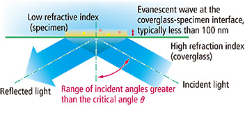
Overview of Evanescent Wave Illumination
CFI Apochromat TIRF series objectives
Numerical apertures of the highest level
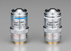
These objectives are the world's first oil-immersion-type objectives to incorporate a correction ring for temperature changes and coverglass thickness. By rotating its correction collar, researchers can easily eliminate spherical aberrations' negative influence on the image quality resulting from temperature-induced changes in the refractive index of the immersion oil and influence from variation in the coverglass thickness. The lenses have been calibrated for a range from 23°C (room temperature) to 37°C (physiological temperature). Additionally, these objectives also provide spectacular images under DIC, epi-fluorescence, confocal, and deconvolution imaging, while providing a strong trapping power during applications using laser tweezers.
SRIC (Surface Reflective Interference Contrast) method can reveal focal contracts prior to switching to TIRF
As evanescent wave illumination excites within approximately 100 nm from the glass surface, TIRF observations require that specimens contact the coverglass, otherwise no TIRF image is obtained. SRIC makes all sections in contact with glass appear black, allowing users to confirm whether a specimen has adhered to the glass before proceeding with TIRF observation. As no excitation light is used in this process, specimen damage is minimized and users can take their time focusing.
Nikon has developed an SRIC system that can be used with both the laser TIRF and white-light TIRF systems. Using SRIC is as simple as switching to the special filter cube.
Laser TIRF image

SRIC image
- Images courtesy of Dr. Masaya Hashido, University of Tokyo