Research Stereo MicroscopeSMZ25/SMZ18
Drosophila embryo dividing timelapse (Epi-fluorescence observation)
(Using SHR Plan Apo 2x, zoom magnification of 8x with SMZ25, GFP; 2.4 hrs total; imaged every 30 secs)
Images courtesy of Max V. Staller, Clarissa Scholes, and Dr. Angela DePace, Harvard Medical School
C. elegans expressing GFP-neurons (Epi-fluorescence observation with OCC illumination)
Single neurons are resolved (in green) above the OCC illumination background. (Using SHR Plan Apo2x, zoom magnification at 3x with SMZ25, GFP and OCC)
Image courtesy of Dr. Julie C. Canman, Columbia University
Zebrafish embryo (Brightfield observation with diascopic illumination)
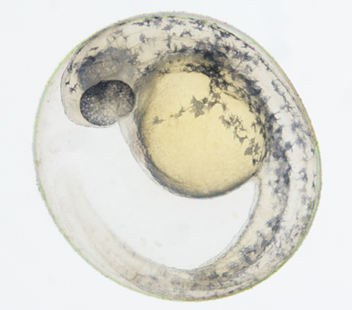
(Using SHR Plan Apo 2x, at zoom magnification of 6x, with SMZ18)
Image courtesy of Junichi Nakai, Ph.D. Saitama University Brain science Institute
Fertilized mouse egg (Epi-fluorescence observation)
The spindle appearing in cell division can be observed
Fertilized mouse egg, Green: Spindle (EGFP- α tubulin), Red: Nucleus (Histone H2B-mRFP1)
(Using SHR Plan Apo 1x at zoom magnification of 13.5x with SMZ25)
Image courtesy of Kazuo Yamagata, Ph.D., Center for Genetic Analysis of Biological Responses, Research Institute for Microbial Diseases, Osaka University
Drosphila brain (Epi-fluorescence observation)
Individual olfactory nerve cells in a drosophila expressing a GFP-membrane marker are clearly resolved as black bodies encircled by fluorescent membranes (see circled area). This image demonstrates the SMZ25's incredible high resolution as the olfactory cells are typically only ø5μm in diameter
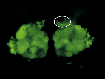
Drosphila brain, GFP-G
(Using SHR Plan Apo 2x at zoom magnification of 15.75x with SMZ25)
Image courtesy of Hokuto Kazama, Ph.D., Laboratory for Circuit Mechanisms of Sensory Perception RIKEN
Zebrafish (Epi-fluorescence observation with OCC illumination)
A single motor neuron expressing clusters of GFP-glycine receptors (resolved as individual puncta along the cell body and processes) imaged in a live zebrafish
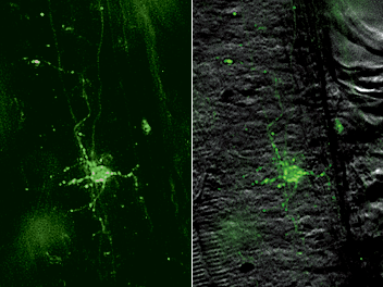
Zebrafish (GFP and OCC)
(Using SHR Plan Apo 2x at zoom magnification of 15.75x with SMZ25)
Image courtesy of Joe Fetcho, Ph.D., Cornell University
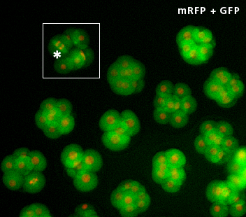
C. elegans embryos (Epi-fluorescence observation)
Time-lapse imaging of developing C. elegans embryos expressing RFP-histones and GFP-membrane markers allows researchers to screen for cytokinesis mutants prior to selection for downstream applications

C. elegans embryos (GFP and RFP; each ovoid is ø30μm in diameter)
(Using SHR Plan Apo 2x at zoom magnification of 8x with SMZ25)
Image courtesy of Julie C. Canman, Ph.D., Columbia University