Upright MicroscopeECLIPSE Ci-E/Ci-L/Ci-S
- Motorized magnification switching
- Bright and uniform Eco-illumination
- Enhanced operational ease
- Effortless image capturing
- Versatile observation techniques
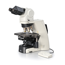
Ci-E Motorized model with LED illumination
Featuring motorized changeover of objective lenses and automatic adjustment of light intensity, ECLIPSE Ci-E has dramatically increased operational efficiency for laboratory tests that require frequent magnification switching. Viewing images, sample changing and capturing images are all conducted with a natural posture. High-luminescent, eco-friendly LED illumination reduces the need for frequent lamp replacement. A variety of accessories are available that support various imaging techniques.
Motorized magnification switching
Magnification can be easily changed during observation with the touch of a button on the ECLIPSE Ci-E microscope body or remote control pad, as well as on the DS-L3 camera control unit's touch panel. Two specific objective lenses can be programmed and switched with the buttons alone. User-defined light intensity for each magnification is automatically saved and reproduced when magnification is changed.

Nosepiece rotation buttons

Remote control pad
Bright and uniform Eco-illumination
The newly developed high-luminescent LED is a low-power-consuming eco-friendly light source that produces evenly distributed illumination and reduces the cost and effort of lamp replacement, thanks to a long life of 60,000 hours. By combining a collimator lens, fly-eye optics and LED illumination, bright and uniform images up to the periphery can be obtained even in high magnification. The LED illuminator features low-heat generation and provides the same color temperature in every magnification
Enhanced operational ease
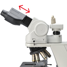
Observation with a natural posture
Using the ergonomic binocular tube, which features an eyepiece that can be inclined from 10° to 30°and extended up to 40 mm, the microscope can be adjusted to suit natural posture. Moreover, a camera can be mounted via the DSC port. The eyelevel riser lifts the eyepiece tube in 25 mm increments (up to 100 mm*) and increases flexibility for more than one user of different eye-point height.
- *Up to 50 mm with ergonomic binocular tube.
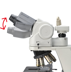
Ergonomic binocular tube

Eyelevel riser
User-friendly stage operation
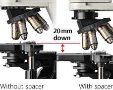
With the addition of a nosepiece spacer, the stage height can be lowered 20 mm from the standard position, facilitating frequent specimen change. The stage handle height can be changed to ensure a comfortable hand position. The stage height can be locked using the refocusing knob, allowing refocusing after specimen changes. The stage is coated with high-durability scratch-resistant ceramic coating.

Height adjustable stage handle
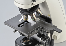
Ceramic-coated stage
Effortless image capturing
One simple click of the image capture button on the microscope base during observation enables the Digital Sight camera to capture the specimen image.
Observation technique icons on the DS-L3 camera control unit's touch panel provide optimal camera settings for each observation method. In addition, ECLIPSE Ci series microscopes can be connected to remote PCs via a DS-L3 to enable remote viewing, online education, and distance collaboration.
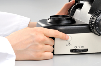
Image capture button
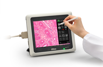
DS-L3 camera control unit
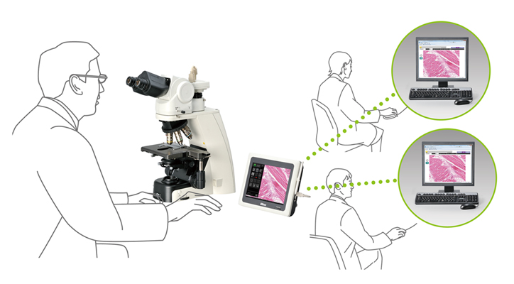
Digital pathology via network
Versatile observation techniques
High-intensity Eco-illumination used in the Ci-E and Ci-L offers sufficient light intensity for phase contrast and simple polarizing microscopy.
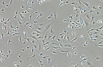
Phase contrast
High-contrast images with neutral background coloration regardless of the magnification range can be captured. This observation technique is suitable for observation of unstained structures.

Phase contrast accessories and objective lenses
Simple polarizing
This is ideal for observing bi-refringent samples such as collagen, amyloids and crystals.
- *Two types of analyzer are available: intermediate tube type and nosepiece slider type.
2.8-Dihydroxyadenine crystals, Simple polarizing, CFI Plan Fluor 40x
Department of Clinical laboratory, Nihon University Itabashi Hospital
Simple polarizing accessories
Sensitive color polarizing
This enables the identification of uric acid crystals forming inside an organism by changing the interference color. It is ideal for gout and pseudo-gout tests.
- *Two types of analyzer are available: intermediate tube type and nosepiece slider type.

Sodium urate crystals, Sensitive color polarizing, CFI Plan Fluor 40x
Department of Clinical laboratory, Nihon University Itabashi Hospital
Sensitive color polarizing accessories
Darkfield

Enables clear observation of blood or the minute structure of flagella. Dry- and oil-type condensers are available. The expander lens is used to capture brighter illumination.
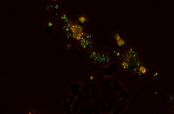
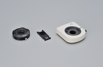
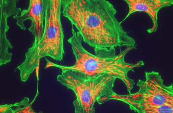
Epi-fluorescence
The compact epi-fluorescence attachment utilizes the dedicated noise terminator mechanism and allows weakly fluorescing specimens to be captured with great clarity and brightness. The filter turret can accommodate up to four filter cubes, and changing them is simple. High-optical-performance objective lenses for epi-fluorescence imaging, including the CFI Plan Apochromat Lambda series and the CFI Plan Fluor series, are available.

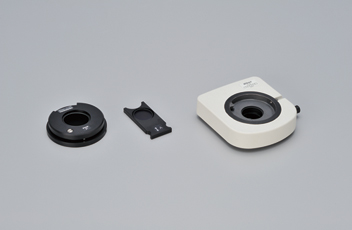
Epi-fluorescence attachment and filter cubes