
1. The ultraminute world becomes the focus of attention in cellular research
The body of a human being engaged in its vital actions consists of multiple systems interacting with one another. The precision of systems such as neurotransmitters and the immune system is nothing short of astounding. The workings of DNA and cells are gradually being revealed, due to the remarkable progress being made in medicine and science. However, many more unseen mysteries still remain. An in-depth understanding of the structures and functions of living cells is required for the elucidation of the mechanisms of vital actions and for efforts to further develop medical treatment, for example through the development of new drugs and the establishment of treatment methods.
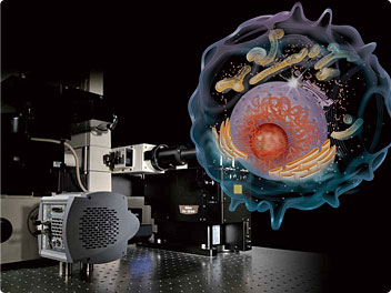
Accordingly, live-cell imaging is now being widely used in cellular research and has shed light on many subjects. With the development of the biosciences in recent years, there is now a greater need than ever for more detailed observation of the minute structures of the cell, such as the membrane and the organelles.
One such feature is an organelle known as the mitochondrion, which plays an important role in support of the vital actions of the cell. Using conventional optical microscopes it has not been possible to visualize the internal structure of mitochondrion in living cells clearly. Electron microscopes, which have higher resolutions than optical microscopes, are used in the examination of the minute structures of cells. However, since this requires the specimen to be frozen and imaged in a vacuum, it is impossible to capture images of live cells using an electron microscope. Thus, although it has been possible to formulate hypotheses based on the results of imaging with an electron microscope, it has been difficult to prove them for living cells.
For reasons such as these, the need has arisen for an optical microscope that can overcome existing limitations and achieve a high degree of resolution.
2. Nearly doubles the resolution of conventional microscopes using moiré patterns
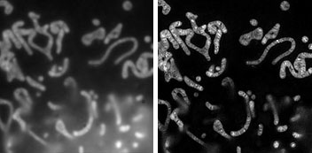
Captured with a conventional microscope (left) and with the N-SIM (right)
The minute structure in the mitochondria can be clearly observed using the N-SIM.
In the context of optical technology such as cameras and microscopes, the term "resolution" signifies the ability to distinguish two close objects as being separate. This is also referred to as "resolving power."
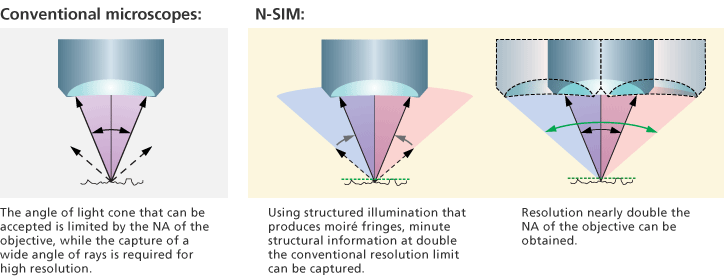
For conventional optical microscopes this resolution has been limited to approximately 200 nm, meaning it is impossible to distinguish two objects that are less than 200 nm apart. To allow this, the objective lens would have to accept a larger angle of the cone of light than that which conventional microscopes can accept. In practice, however, there are theoretical limits to the light acceptance angle of an objective lens, which is restricted by the NA of the objective lens and the wavelength of the light.
Super Resolution Microscope N-SIM has overcome the limitations of the optical microscope, achieving a spectacularly improved resolution of nearly double that of conventional microscopes. The N-SIM is an optical microscope that achieves super resolution by combining proprietary Nikon optical technology with Structured Illumination Microscopy (SIM), a technique that uses the moiré phenomenon to increase resolution and which was announced by a team led by Dr. Mats G. L. Gustafsson of the University of California, San Francisco, in 2000.
- *Group leader (2008—2011), Janelia Farm Research Campus, Howard Hughes Medical Institute

The moiré phenomenon
Moiré patterns are striped patterns caused by interference. This phenomenon occurs when multiple regular patterns are superimposed on top of one another in such a way as to produce a pattern that differs from the original pattern. When a finely striped pattern (structured illumination) is applied to a specimen, moiré patterns are produced. Due to the fact that the moiré patterns generated are characteristically coarser than the original pattern, it is possible to capture them with an optical microscope. The captured moiré fringes contain minute structural information of specimens. As a result, it is possible to mathematically restore the sub-resolution structure of a specimen by analytical processing of multiple moiré images captured while slightly varying the orientation and phase of the structured illumination.
The adoption by the N-SIM of this groundbreaking technology enables it to achieve significant resolving power using a new illumination scheme without significant alteration of the structure of the microscope.
3. Enables super-resolution time-lapse imaging of live cells
In order to meet the requirements of live-cell imaging, it is not enough simply to increase resolution; it is also important to achieve sufficient speed so as to capture dynamic molecular interactions in living cells. However, it is no simple matter to reduce the processing time for structured illumination microscopy, in which a single super-resolution image is constructed out of multiple images.
Nikon exhaustively pursued shorter image acquisition times to visualize dynamic behavior in living specimens. Nikon proceeded to work on improved objective-lens performance and image-processing techniques, as well as on the development of high-precision optical systems for structured illumination that would generate moiré fringes effectively. As a result, clear super-resolution time-lapse images are successfully captured as fast as 1.67fps, as only nine images are required for the construction of a single super-resolution image*.
- *With 2D-SIM mode
By focusing on structured illumination microscopy and integrating unique Nikon technologies, Nikon has created the N-SIM, which enables super-resolution live-cell imaging. It is anticipated that this microscope will help overcome various challenges in today's bio-imaging field and greatly expand the realm of cellular research.
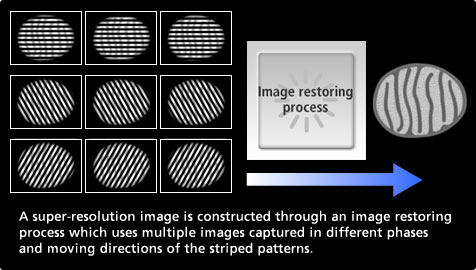
Since the N-SIM can construct a super-resolution image using as few as nine images (three directions, three phases) in 2D-SIM mode, and as few as fifteen images (three directions, five phases) in 3D-SIM mode, the image acquisition time is correspondingly short.
Fast time-lapse imaging of mitochondria with the N-SIM.The N-SIM is the first microscope in the world to successfully capture clear and fast time-lapse images of the pleated structures known as cristae in living mitochondria. (This movie clip is a loop sequence which repeats the recording of these mitochondria five times.)