
1. Regenerative medicine, the rising star of advanced medical treatment

Master cell research is rapidly gaining interest in Regenerative Medicine.
Regenerative medicine, also known as tissue engineering, is a rapidly growing multi disciplinary field attracting worldwide attention as a major step in advanced medical treatment. Regenerative medicine refers to developing functional cell, tissue, and organ substitutes to repair, replace or enhance biological functions that have been lost due to congenital abnormalities, injury, disease, or aging.
Until now, regenerative medicine had primarily focused on research into ES (embryonic stem) cells *1. Human cells in the brain, liver, muscles and other tissues are different from one another; while ES cells can be altered into any type of cell and are thus referred to as master cells. ES cells are derived from human embryos and utilizing these cells for research have caused significant ethical concerns.
- *1Embryo stem (ES) cells: Fertilized mice embryos cells develop into placenta and other tissues in three to five days following fertilization; and there are cell masses that develop into the mouse's body itself. ES cells are established as undifferentiated cell lines in culture outside the mouse's body. ES cells have the ability to transform into various types of cells. Human ES cells were first successfully cultured in 1998.
IPS (induced pluripotent stem) cells are produced by inducing skin cells to revert back into original master cells through a process of activating certain genes. *2. For example, cells can be taken from a patient's skin, turned into iPS cells, and after they are transformed into tissue cells, researchers believe it is possible to make 3D structures which organs are made from. Because iPS cells are made from the patient's own skin cells, this process does not face the rejection issue that can occur with organ transplantation. Additionally since iPS cells are made from skin cells, there are no ethical dilemmas as there are with ES cells.
While research into the practical use of iPS cells in medical treatment is continuing, in the area of new drug therapy the use of muscle and liver cells made from iPS cells in drug trial efficacy and side effects is promising.
- *2Induced pluripotent stem (iPS) cells: IPS cells have been given properties similar to ES cells by introducing various types of genes (transcription factors) to somatic cells (mainly fibroblastic cells). These cells were first produced by a research group led by Professor Shinya Yamanaka of Kyoto University.
2. BioStation CT, providing a new style of research instrument
IPS cell research is said to have an extremely low success rate. Transformed skin cells can regress to being master cells and little is known about the cellular mechanism that cause master cells to change to tissue cells.
To understand this mechanism, it is necessary to continually observe the cells while they are alive. Such observations are difficult, however, because living cells do not tolerate even slight changes in their environment well. A safe methodology for detailed observations of living cells under consistent conditions is a key for iPS cell research and regeneration medical laboratories that grow these cells. The Nikon BioStation CT represents a viable solution to this issue.
Is research in living cells really that difficult? In the world of bioscience, live cell research require incubators that mimic the conditions that normally exist inside the living organism. (37°C, 95% humidity, and 5% CO2 concentration. During the growing prccess; there are times when cells need to be removed from the incubator to be examined microscopically. This is tedious work and during this short observation time, cell activity can diminish and cells can become contaminated.
The BioStation CT reverses conventional ideas with its innovative approach of mounting a microscope inside the incubator. As a result, live cells can be observed while they are being cultured and samples do not need to be removed. And since a digital camera can be mounted on the microscope, images of the observations can be captured continuously and added to a database.
As with private enterprises, the concept of "automation" has recently gained prevalence at research organizations for the sake of greater efficiency and increased throughput. The Nikon BioStation CT can be considered the first product to achieve significant cell culture laboratory automation. While its use is still experimental, it is already being used in iPS cell and cancer cell research with promising results.
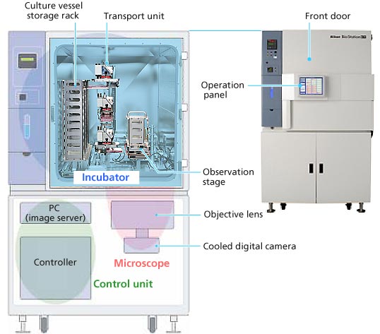
BioStation CT configuration
Observation of live cell cultures with the BioStation CT
3. Nikon technology that makes the BioStation CT possible
Cells are transparent and colorless. Nikon technology is well suited to observing these transparent, colorless cells. The BioStation CT uses Nikon's proprietary APC (Apodized Phase Contrast) objective lens. The phase-difference lens that emphasizes contrast using the refractive index variance of material inside the cells has been developed to enable the observation of cells in greater detail, providing documentation of cells during division. Such observation is difficult with conventional phase-difference lenses.
Principle of phase-difference observation
The APC objective uses the nature of diffracted light *3 to widen the angle at which it passes through a tiny cell. Cell detail is made more visible by inserting absorption bands of different transmittances inside and outside the phase ring in the lens. The halo*4 that appears around the cell makes the cell look brighter if it is darker than the background, or if it is bright, it makes it look much brighter than the background.
With the current BioStation CT, researchers check the condition of samples through the monitor, but Nikon is now developing a new feature that combines Nikon microscope and image analysis technologies to automatically observe close-up changes in cells or time replacement of culture media *5. Nikon is also evaluating incorporating a robot for changing culture media. When this new design is realized, automated production of live cell cultures will advance further, enhancing efficiency in regenerative medical treatments and new drug therapy research.
- *3Diffracted light: Light that bends around to the rear of the cell
- *4Halo: a circle of light surrounding a cell
- *5Culture medium: Fluid containing nutrients required for the growth of bacteria or cells
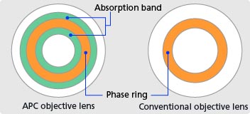
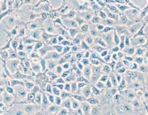
Image observed with conventional objective lens
Halo appears at the boundary section of the cell.
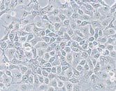
Image observed with APC objective lens
Halo fades and can be observed up to the detailed section.