
Normally, the cells that make up our bodies are not transparent. These cells contain substances that perform various functions, and these substances emit color when they absorb light. However, transparent tissue does exist within our bodies—the lenses of our eyes. The two key factors in the transparency of the lens of the eye are the transparency of the cells that comprise it and the regular configuration in which these cells are arranged.
Permanently frozen ice
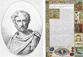
Portrait of Pliny the Elder and the Naturalis Historia, published in 1472
© Giraudon/PPS
Pliny the Elder of ancient Rome described crystals as “permanently frozen ice.” Rome itself was decorated with transparent crystals to help keep people cool during the summer.
As well as being a natural historian, Pliny the Elder was also a statesman and soldier. His masterwork was his 37-volume Naturalis Historia, which covered a wide range of subjects, including geography, astronomy, botany and mineralogy. With its descriptions of monsters and giants, there are parts of this work that are of questionable scientific value; however, as a compilation of contemporary knowledge, it represents a major achievement. Crystals are of course not permanently frozen ice, as they are described in the Naturalis Historia; however, the fact that Pliny’s description of this transparent mineral, which could be found everywhere, survives is extremely interesting in itself. With the emergence of printing technology in the Middle Ages, the Naturalis Historia was published and it has continued to be read ever since.
As a common mineral, crystal has been well known since ancient times. More than anything else, people have probably been enchanted by its high degree of transparency.
In fact, our bodies contain tissue that is transparent like crystal. If all our tissue were transparent, we would probably be invisible. Normally, however, tissue is colored. The transparent structure in our bodies that functions as a lens is known as the “crystalline lens.”
Transparent crystals
The transparent tissue in the eye is known as the “crystalline lens.” As the name suggests, the role of this tissue is to focus the eye; when we look at an object that is close, the muscle that supports the crystalline lens makes it thicker and, conversely, when we look at a distant object, the muscle acts to make it thinner.
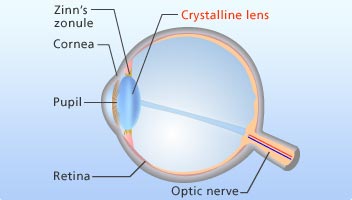
The structure of the eye: Zinn’s zonule adjusts the thickness of the crystalline lens to focus the eye, while the crystalline lens is nourished by the aqueous fluid that is present between the cornea and the crystalline lens.
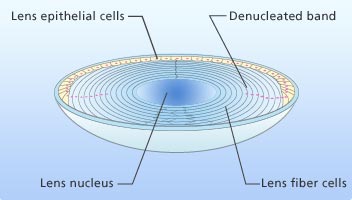
The structure of the crystalline lens
Within the human body, the crystalline lens has the distinction of being transparent tissue.
The crystalline lens resembles an onion in structure, with approximately 1,000 layers of cells arrayed with extreme regularity. Two factors are keys to preserving the transparency of the crystalline lens: first, that there is no excess matter contained in the cells that could alter the refractive index within the cell, and second, that the cells themselves are aligned in a regular fashion so that they do not alter the refractive index. Although the cells cannot be said to be perfectly transparent, since the cell walls slightly disperse light, they do, in the main, preserve the transparency of the crystalline lens.
Surprisingly, the cells that comprise the crystalline lens are never replaced over the period of an individual’s life. Most of the body’s tissue cells are constantly replaced by new ones—with approximately 90% of cells replaced every six months. However, with the crystalline lens, especially its center portion, the same cells are used throughout one’s entire life.
The fact that new cells cannot be generated means that the crystalline lens is unable to repair itself if it is damaged. Cataracts (as seen in many elderly people) are caused by a gradual clouding of the crystalline lenses over many years. The only way of treating this is to surgically replace the opacified crystalline lens with an artificial one.
Why, then, are new cells not produced in the crystalline lens, thus making it unable to repair itself? The answer to this question is related to the transparency of the cells.
Focusing on the natural death of cells
Ordinary cells contain various organelles, such as DNA—the blueprint for life. If these organelles are present, however, they alter the refractive index—making the cells non-transparent. It is because the cells in the crystalline lens do not contain these organelles that they preserve their transparency. Since cells that do not contain organelles also lack the cellular blueprint of DNA, they are unable to generate new cells and carry out repairs.
Organelles such as DNA are present when the cells of the crystalline lens are being developed, before birth. However, the organelles are destroyed immediately after we are born.
How and why are the organelles of the crystalline lens destroyed? In an effort to shed light on this process, the focus has been on the mechanism of “cell death.”
In addition to “apoptosis,” or programmed cell death, which occurs in cells throughout the entire body, and which allows old cells to be replaced with new ones, there are two other types of cell death: “necrosis,” in which part of the body dies, and “autophagy,” in which cells consume themselves. In contrast to necrosis, which is caused by infection or a decrease in blood flow, the occurrence of apoptosis enables the renewal of the body’s cells. When apoptosis occurs, the targeted cell is broken down into small pieces; normally, once apoptosis has started, it does not stop until completion.
Recent research points to apoptosis being extremely well controlled at the time that the crystalline lens is created, and that although the organelles are broken down, the apoptosis functions in such a way that the outer cell wall, the cell’s cytoskeleton, and the crystallin proteins remain intact. The true picture remains unclear, however, since there is also research that suggests that the lack of organelles in the crystalline lens is the result of autophagy.
From a left-handed form to a right-handed form

L-type at left, D-type at right. When the amino acid in the protein changes to D-type, the structure is thrown into disarray.
Even in a crystalline lens that has maintained its transparency, the onset of aging brings on the turbidity known as a cataract. This is due to crumbling protein, which becomes cloudy and interacts poorly with other crystallins. Although cataracts have been known for a long time, it is only recently that the change in the structure of amino acids has come to light—one reason for the crumbling protein.
Incidentally, amino acids, which are constituents of the proteins that make up living organisms, come in two forms: “right-handed” or “D-type,” and “left-handed” or “L-type.” D-type and L-type amino acids have the same chemical properties, but are the mirror images of one another. Incredibly, all living organisms include L-type amino acids. There is not a single living organism that is made up of proteins that consist solely of D-type amino acids. This is yet another biological mystery.
Some proteins in some living organisms contain D-type amino acids, and in 1978 it was discovered that D-type amino acids in the crystalline lens increase with the onset of aging. By studying the crystalline lens in detail, a research group led by Professor Noriko Fujii of Kyoto University ascertained that the aspartate residue (a type of amino acid) at one particular site in the crystalline lens is inverted to D-type.
The group discovered that when D-type amino acids are formed in proteins, the structure of the proteins around the D-type amino acids are distorted, as a result of which the crystallins will aggregate (or clump together) in an abnormal fashion. If these aggregates are larger than the wavelength of light, whitish, cloudy cataracts are formed. L-type amino acids in proteins inverted to D-type as aging progresses has also been seen in cases of Alzheimer’s disease and Parkinson’s disease.
Professor Fujii’s research group is now looking into why the aspartate residue in proteins is inverted from L-type to D-type. Other amino acids do not normally make this change in the benign environment of a living organism; however, due to the peculiarity of its side chain (a chemical group that is attached to a core part of a molecule), aspartate residue has a property that makes it prone to changing from L-type to D-type. The group also discovered which locations within the proteins make the aspartate residue prone to this inversion. It has also become clear that this phenomenon is accelerated by exposure to ultraviolet light.
The transparency of the crystalline lens is maintained by a combination of two factors: the mechanism that keeps the cells themselves transparent, and the preservation of a regular cell structure. What is surprising is that the mechanism is so clever. Delving into why the crystalline lens remains a crystalline lens is shedding light on a fundamental mechanism of living organisms.
Cooperation on materials and research / Date of article posted
Professor Noriko Fujii, Radiation Biochemistry and Biological Function,
Division of Radiation Life Science Research Reactor Institute, Kyoto University
Associate Professor Hiroyuki Matsushima, Department of Ophthalmology, Graduate School of Medicine, Dokkyo Medical University
Saori Sakagami and Hirofumi Seo, Faculty of Medicine, University of Tokyo / August 2010