Shear amount
Differential interference is a phenomenon used to observe contrast in specimens such as extremely fine structures or transparent biological specimens (phase specimens) for which conventional human sight or microscope cameras cannot discern contrast. Differential interference contrast (DIC) microscopy involves optical techniques not used in brightfield microscopy, the most important of which is a special crystal prism in the light path.
In conventional brightfield microscopy, light passes through the objective to bounce off the specimen and back through the objective to form the image. In DIC microscopy, a special crystal prism is inserted before the objective (Figure 1). This has the following effect:
- The prism splits the light into two paths.
- The light paths split by the prism diverge by a very slight amount, called a “shear amount”. These split light rays will bounce off the specimen at different points, separated only by the shear amount, and back through the object lens to the viewer.
- Light rays passing back through the objective overlap in the prism. The spatial difference between the points at which the two light paths reflected off the specimen result in an optical path difference (phase difference) which results in interference when they overlap again within the prism, imparting contrast equivalent to the difference of the light paths.
The spatial difference between the points at which the two light paths reflected off the specimen result in an optical path difference (phase difference) which results in interference when they overlap again within the prism, imparting contrast equivalent to the difference of the light paths.
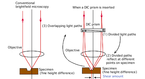
Figure 1. Shear amount in differential interference (reflective) microscopy
This technique allows contrast that would be nearly invisible in brightfield microscopy to be viewed in DIC microscopy, lending high contrast sensitivity to phase specimens that are difficult to view. However, resulting images have directionality, and contrast is visualized only in the direction that the light paths diverge. This is called the “shear direction”.
- For more information
-
-
 Microscopy U
![[Open in a new window]](/common/img/icon/window.png)
The source for microscopy education.
-