Microscopy imaging techniques
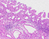
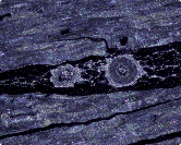
The most commonly used microscopy imaging technique is brightfield microscopy, where light is either passed through or reflected off a specimen. However, there are also techniques known as darkfield microscopy, phase contrast microscopy, differential interference contrast (DIC) microscopy, fluorescence microscopy, and polarizing microscopy. Each of these methods is best suited for different uses. Research class microscopes allow these techniques to be utilized merely by switching attachments.
Brightfield microscopy
Observation is made by viewing the light passed through or reflected off a specimen.
Darkfield microscopy
A special condenser lens is used to illuminate the specimen diagonally, then observe light scattering off it. The field of view is darker than brightfield microscopy because illumination light does not enter the objective lens.
Main uses:
- Microbiological imaging
- Blood tests
- Detecting microscopic scratches or irregularities
Phase contrast microscopy
The optical phenomena of diffraction and interference are used to add light/dark contrast to a transparent specimen for imaging. There is no need to stain the specimen as in brightfield microscopy, so live specimens can be used.
Differential interference contrast (DIC) microscopy
Differential interference contrast microscopy transforms minute differences in refraction indexes of light passing through an unstained specimen, or optical path differences from the specimen surface shape, into a monochromatic shadow-cast image enabling observation. As with phase contrast microscopy, DIC microscopy may be used with living specimens. However, it is better suited to thicker specimens.
Main uses:
- Imaging fibrous structure of nerve or muscle
- Imaging mitotic spindles
- Imaging cellular nucleic structures or other thick unstained specimens
- External examination of IC wafer surfaces or polished surfaces of magnetic heads
- Observation of crystal growth process
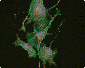
Fluorescence microscopy
Specimen is excited with a specific wavelength of light, then fluorescent emanations are observed.
Main uses:
- Imaging and quantification on calcium ion dynamics in living cells
- Assaying antigens in antigen/antibody reactions
- Imaging and quantification of intracellular DNA and RNA
- Analysis of chromosomal abnormalities
- Detection of resists remaining in semiconductor fabrication process
- Detection and assaying of foreign particles
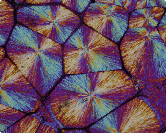
Polarizing microscopy
This technique uses the phenomenon of polarization to add contrast and color to specimen images.
Main uses:
- Analysis of optical properties of rocks, ores, polymers, and experimental materials
- Detection of defects in the liquid crystal display element fabrication process
- Detection of defects in separation membranes
- Imaging magneto-optic disks and bubble memory
- Polarization analysis of fine structures within living organisms and cytoskeletons
- Gout testing

